
MS Thesis and Research
Thesis Title:
Identification of relationships between extracellular matrix and aneurysmal mechanism
Expected Completion: April 2018
Big Picture
Background
Intracranial aneurysms are pathological enlargements of the walls of cerebral arteries, estimated to exist in approximately 5% of the adult population. As part of my MS thesis,
I am researching about the mechanobiology that affects and governs their development, enlargement, as well as rupture. Solving the aneurysm puzzle is an engineering one, much like calculating when a damaged building will likely collapse. My primary interest lies in understanding how the heterogeneity found in cellular architecture correlates with the mechanical properties observed from a cerebral aneurysm wall.
The ultimate goal is to develop a formula or algorithm that physicians could use to more accurately determine the rupture risk.
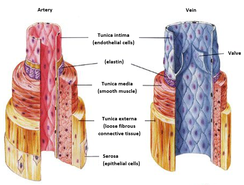
In a brief overview, collagen and smooth muscle cells are the key elements that maintain the wall’s structural integrity, while the elastin layer facilitates its flexibility to accommodate pulsatile blood flow. In a large percentage of aneurysms,
the endothelium is missing, which is the inner-most layer that protects the arterial wall from being directly in contact with blood. Conventionally, we think that without such protection, inflammation takes place, causing subsequent disturbances in the adjacent arterial layers and mural contents.
Yet, because of the unpredictable nature associated with rupture, it has been a challenge to fully examine the transitional change in pathology, which occurs at the immediate instants before and after rupture takes place. For this reason, what we know so far remains a conjecture.
Furthermore, during assessments on the aneurysmal wall’s strength, there is a fundamental gap in our knowledge about the mechanism, by which the abnormal hemodynamics within the aneurysm sac can lead to a breakdown in the normal process of collagen renewal and remodeling.
Consequently, under hemodynamic stress, an aneurysm would be left vulnerable to mechanical failure. There is no single or direct indicator that can precisely predict the likelihood of such failure. Although aneurysm size is widely used as a measure for evaluating rupture risk,
the ultimate size at time of rupture varies drastically between individuals.
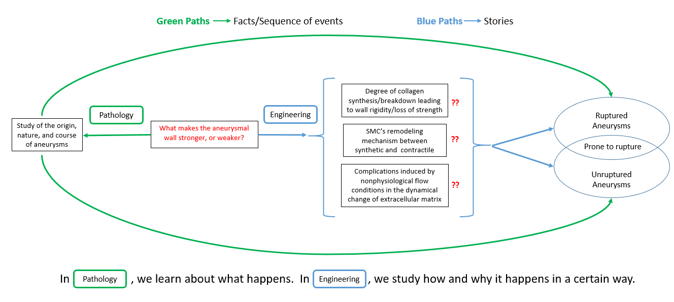
To address the unknowns, a major focus of engineering studies done in my research group is on improved risk stratification via using a combination of measurable clinical parameters, such as histological studies, aneurysm flow dynamics, mechanical tests, and aneurysm geometry.
Research Goals:
At a high level, the ongoing investigations are summarized as follows,
1. Obtain relationship between aneurysmal wall thickening and proliferation of cells.
2. Identify the combinatorial patterns and distribution of cellular materials in aneurysmal wall.
3. Study the relevant mechanical testing to find linkage with histological results.
4. Elucidate the physics behind the observed kinematic change in cerebral aneurysm wall.
5. Interpret findings by comparing with other literatures
Structure of an artery
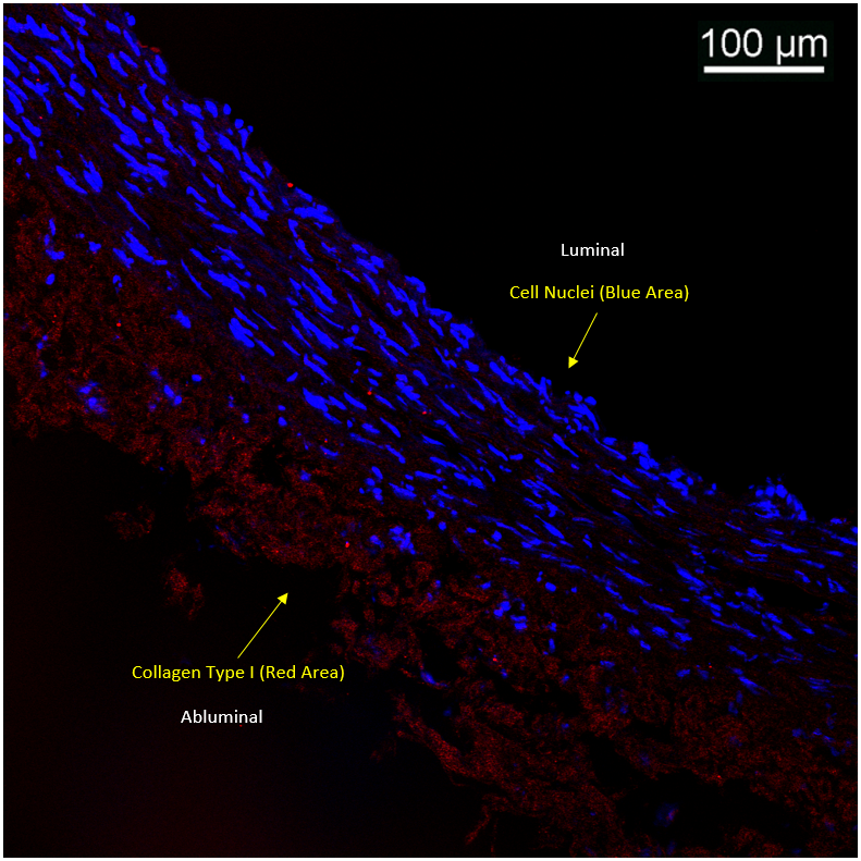
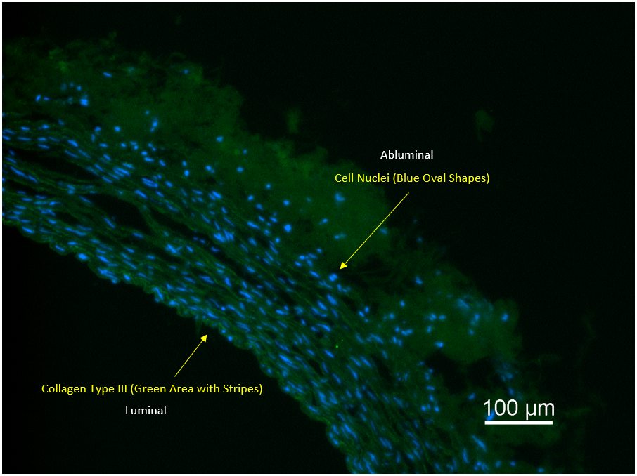
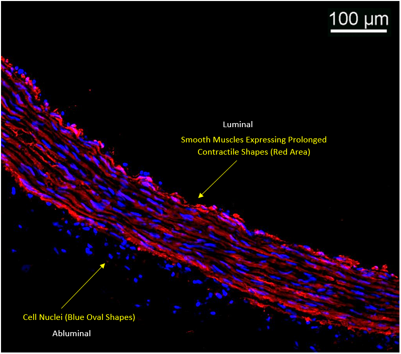
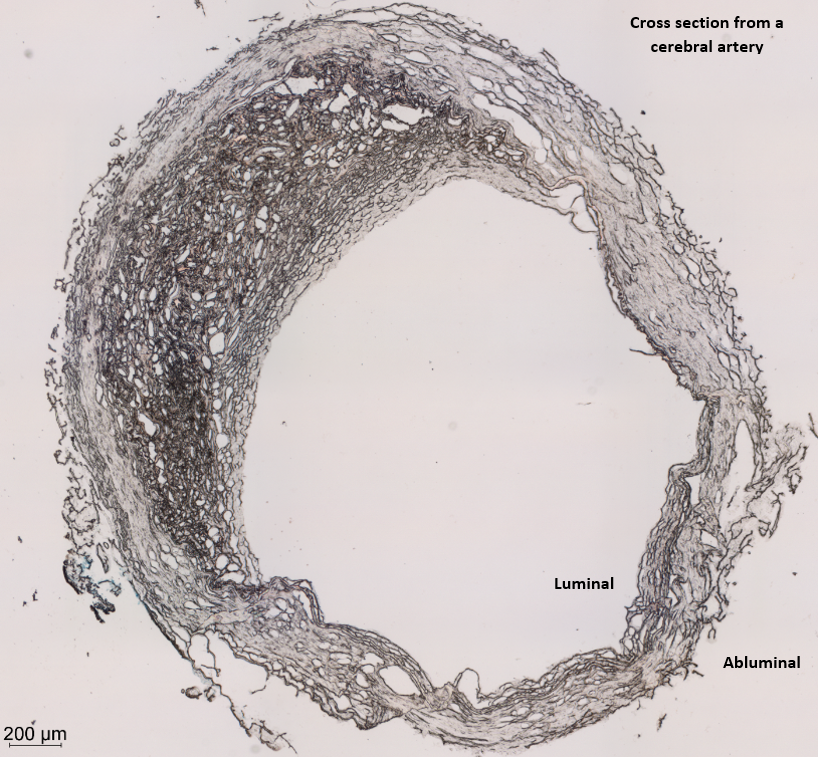
Histology Staining / Studies on Human Cerebral Artery:
Background Information:
Cross sections were cut, using cryostat, from a segment dissected from the cadaver Circle of Willis. Relevant protocols were applied to stain cells nuclei, collagen type I,
collagen type III, and smooth muscles, the results of which can be seen in the below figures. Also included is a cross section image taken by bright field microscopy. Histological studies are important because they can
help us understand in details the encoded messages behind pathological mechanisms, which can not be easily visualized.
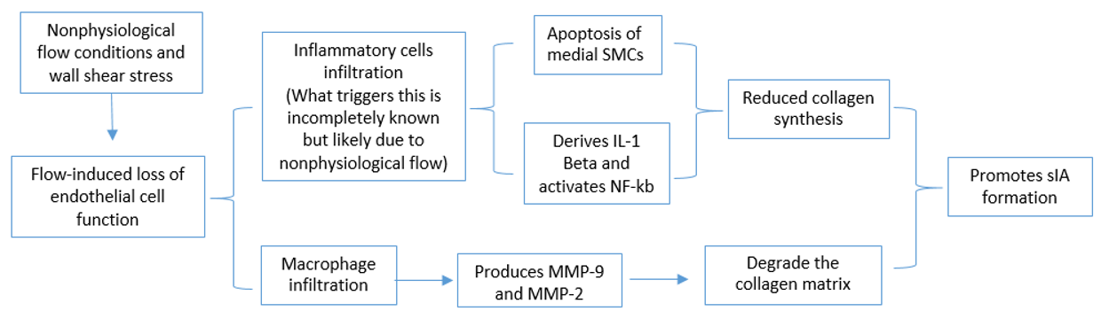
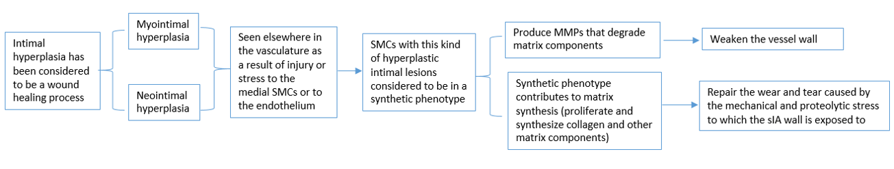
Conjectured Models of Arterial Aneurysms:
Nonphysiological Flow Conditions:
Intimal hyperplasia:
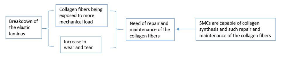
Breakdown of Elastic Lamina:
Reference for Picture (Structure of an artery):
Weber, Craig, MD. Comparison of Arteries (left) & Veins (right). Digital image. The Role of Arteries in the Circulatory System. N.p., Aug.-Sept. 2017. Web. https://www.verywell.com/part-2-arteries-1763959.